Tuesday, January 25, 2022

Tibia bone.

INTRODUCTION

- Medial bone of the leg.
- Stronger bone of the leg.
- Takes part in formation of the knee and ankle joints and thereby helps jn transmission of body weight from femur to the foot.

ANATOMICAL POSITION

- Upper end is broader.
- Anterior border of the shaft is sharp and prominent.
- Medial malleolus projects from medial side of lower end.

PARTS

1. Upper end

Has:-

a. Medial condyl - articulate with medial condyl of femur and has four surface - anterior, medial, lateral, posterior.

b. Lateral condyl - articulate with head of fibula.

c. Tuberosity of tibia - 
has two parts namely :- upper smooth and lower rough part.

2. Shaft
Has -

a. Three borders 
- Anterior, medial and interosseous

b. Three surfaces 
- Medial, lateral and posterior

3. Lower end 
Has -

a. A process known as medial malleolus.
b. Five surfaces (anterior, medial, posterior, lateral and inferior)

ATTACHMENTS

a. Muscles
Origin

1. Tibialis anterior 
2. Soleus
3. Flexor digitorum longus
4. Tibialis posterior

Insertion
1. Sartorius
2. Semi membranosus
3. Gracilis
4. Semi tendinosus
5. Popliteus

b. Ligaments

1. Capsular ligament of knee joint.
2. Deeper fibres of tibial collateral ligament.
3. Capsular ligament of superior tibiofibular joint.
4. Tendon of popliteus.
5. Anterior cruciate ligament.
6. Posterior cruciate ligament.
7. Tibial collateral ligament.
8. Deltoid ligament.
9. Interosseous tibiofibular ligament.
10. Capsular ligament of ankle joint.
11. Ligamentum patellae.

c. Meniscus

1. Posterior horn of lateral meniscus.
2. Anterior horn of lateral meniscus.
3. Posterior horn of medial meniscus
4. Anterior horn of the medial meniscus.

d. Other structures

1. Medial patellar retinaculum.
2. Superior extensor retinaculum.
3. Iliotibial tract.

CLINICAL ANATOMY 

1. Lower one third of tibia has a comparatively poor blood supply, so non union fracture is common in the lower end of tibia.
2. Inversion injury results in transverse fracture of medial malleolus at base.
3. Upper end of tibia is one of the commonest sites for one osteomyeletis


RELATIONS - 
Nerve 
1. Deep peroneal nerve.
Blood supply
2. Posterior tibial artery.

Fibula bone.

It is the lateral and smaller bone of leg and is homologous with ulna of the upper limb.


ANATOMICAL POSITION

Upper end/head is slightly expanded in all directions. The lower end or lateral malleolus is expanded antero posteriorly and is flattened from side to side.

PARTS 

1. Upper end - 
- Articulate with lateral condyle of tibia.
- Have neck below the neck.

2. Shaft - 
- Three borders namely, anterior, posterior, interosseous.
- Three surfaces namely medial, lateral and posterior.

3. Lower end 
- Four surfaces namely, anterior, posterior, lateral and medial surface.

ATTACHMENTS 

a. Muscles 
Origin

1. Extensor digitorum longus.
2. Extensor hallucis longus.
3. Peroneus tertius.
4. Peronius longus.
5. Peronius brevis.
6. Soleus.
7. Flexor hallucis longus.
8. Tibialis posterior.

Insertion
1. Biceps femoris.

b. Ligaments

1. Fibular collateral ligament.
2. Capsular ligaments of superior  tibiofibular joint. 
3. Anterior intermuscular septum.
4. Superior extensor reticulum.
5. Superior peroneal retinaculum.
6. Posterior intermuscular septum.
7. Interosseous membrane. 
8. Interosseous tibiofibular ligament.
9. Anterior tibiofibular ligament.
10. Posterior tibiofibular ligament.
11. Anterior talo fibular ligament.
12. Inferior transverse tibiofibular ligament
13. Calcaneofibular ligament.
14. Posterior talo fibular ligament.

CAPSULE 
1. Capsule of ankle joint.

NOTE - Tendon of peroneus brevis and peronius longus.

CLINICAL ANATOMY 

- Fibula bone is used in bone grafting.
- Lateral malleolus and the ligaments attached to it are very important in maintaining stability of ankle joint.
- Upper and lower ends of fibula are subcutaneous and palpable.

Note - 
1. Spiral fracture of lateral malleolus in first degree of pott's fracture.
2. Fracture of medial malleolus in second degree of pott's fracture.
3. Posterior margin of lower end of tibia in third degree of pott's fracture.

- Forcible abduction and external rotation of foot causes a spiral fracture of lateral malleolus and then fracture of the medial malleolus.


Monday, January 24, 2022

Patella bone

INTRODUCTION
- Largest sesamoid bone.
- Developed in the tendon of the quadriceps femoris

ANATOMICAL POSITION

- Anterior rough surface is placed anteriorly with its apex pointing downwards.
- Posterior articular area is placed posteriorly.

SIDE DETERMINATION

- Triangle in shape with its apex downwards(apex is non-articular structure in patella which lies inferiorly).
- Posterior articular surface is divided by a vertical ridge into larger lateral and a smaller medial areas.
- When bone is laid on the table, it rests on broad lateral articular area which determine its side.

PARTS 

- Has an apex, three borders (superior, lateral and medial) and two surfaces (anterior and posterior).

NOTE :- During various phases of movements of the knee, different portions of patella articulates during extention. Middle pair during beginning of flexion. Upper pair during midflexion. Medial strip during full extention of knee.

ATTACHMENTS

A. Muscles (only insertion)

1. Rectus femoris
2. Vastus intermedius
3. Vastus lateralis
4. Vastus medialis
These four muscles  are called quadriceps femoris.

CLINICAL ANATOMY

1. Patella may get fractured.
2. Bursitis occurs in prepatellar and subcutaneous infrapatellar bursae.

RELATIONS

1. Tibial nerve
2. Common peroneal nerve
3. Fibular nerve


Saturday, January 22, 2022

Femur bone.

Also known as thigh bone and is the strongest bone.

SIDE DETERMINATION

1. Upper end bears a rounded head where      as lower end js widely expanded to
    form two large condyles.
2. Head is directed medially.
3. Cylindrical shaft us convex forewards.

ANATOMICAL POSITION

1. Head is directed medially upwards and      slightly forewards. 
2. The shaft is directed obliquely                       downwards and medially so that the           lower surfaces of two condyles of               femur lie in the same horizantal plane.

PARTS 

1. Upper end 
a. Head
b. Neck - makes an angle if 145° in adult         with the shaft.
Note - Neck has two borders and two                     surfaces named, upper and lower                 border and anterior and posterior                 surface.
Angle of femoral torsion or angle of anteversion is formed between the transverse axis of the upper and lower end of femur. It is about 15°.

2. Shaft
- It is convex foreward.
- Middle one - third of shaft has medial,         lateral and posterior border and anterior,     medial and lateral border.
- Upper one third has 
  4 borders - medial, lateral, spiral line and     lateral hip of the gluteal tuberosity.
  4 surfaces - anterior, medial, lateral and       posterior. 
- Lower one - third of shaft has 4 borders      named medial, lateral, medial                        supracondylarline and lateral supra              condylar line.

3. Lower end

Have two condyles named lateral and medial condyle and intercondylar notch is present between two condyles.

Articular surface - 
Two condyles articulates with patella and parts.

ATTACHMENTS

a. Muscles.

Insertion muscles. 
1. Piriformis
2. Gluteus minimus
3. Obturator internus
4. Obturator externus
5. Gluteus medius
6. Psoas major
7.  Iliacus
8. Adductor magnus
9. Ouadratus femoris
10. Gluteus maximus
11. Adductor longus
12. Adductor brevis
13. Pectineus
14. Two gemelli
15. Ischial head of adductor magnus

Origin muscles
1. Medial head of gastrocnemius
2. Vastus intermedius
3. Articularis genu
4. Vastus lateralis
5. Vastus medialis
6. Biceps femoris
7. Plantaris
8. Lateral head of gastrocnemius
9. Popliteus

b. Ligaments

1. Round ligament 
2. Capsular ligament
3. Iliofemoral ligament(upper band)
4. Iliofemoral ligament(lower band)
5. Medial and lateral intermuscular septa
6. Fibular collateral ligament
7. Tobial collateral ligament
8. Anterior cruciate ligament
9. Posterior cruciate ligament
10. Oblique popliteal ligament

REALTIONS

1. Nerves

a. Femoral nerve
b. Sciatic nerve

2. Blood supply

a. Lateral epiphyseal branch of medial            femoral circumflex artery.
b. Obturator artery.

CLINICAL ANATOMY

- Tripping over minor obstructions causing     forced medial rotation of the thigh and       leg during the fall results in :- 

1. Fracture in shaft of femur.
2. Bucket handle tear of medial meniscus.
3. Pott's fracture of leg bones.
4. Fracture in neck of femur which can            leads to avascular necrosis of head.     

     Note : - If Q angle is increased then             there maybe lateral and subluxation if          patella.


Sunday, January 9, 2022

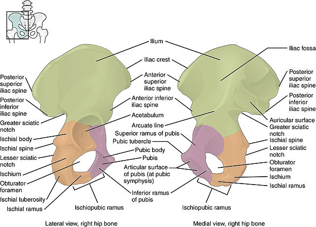
Hip bone

It is a irregular bone present in the hip region of the body which is expanded above and below but constricted in the middle.

Articulations - Hip joint and pubic symphysis.

There are three parts of hip bone named ilium which is present superiorly, ischium present posteroinferiorly and at last pubis which is present antero inferiorly.

ANATOMICAL POSITION
1. Pubis should faces forewards and medially above obturator foramen. 
2. pubic tubercle and anterior iliac spne in same plane.
3. symphyseal surface of pubis lies medially.
4. ilium is expanded and projects upwards.

SIDE DETERMINATION
1. Acetabulum lies laterally and ilium lies above acetabulum.
2. Direction of acetabulm will determine the side.

ILIUM
~ Upper part of hip bone.
~ 2/5th part of acetabulum is formed by ilium.
~ Flat and expanded portion above acetabulum cavity.
 ~ Upper end expanded and known as iliac crest.
~ Lower end is smaller and fused with pubis and ischium and has iliac tuberosity.
~ Three borders named anterior, posterior and medial border is present in ilium.
~ Along with three borders, three surfaces are also present known as gluteal, iliac fossa and sacropelvic.

Note :- Sacropelvic surface has iliac tuberosity.

ATTACHMENTS 

1. MUSCLES

a. Sartorius
b. Fascia lata
c. Tensor fascia lata
d. External oblique abdominis (insertion).
e. Lattisimus dorsi
f. Transversus abdominis
g. Quadratus lamborum
h. Lumbar fascia
i. Internal oblique abdominis
j. Gluteal maximus
k. Erector spinae
l. Piriformis
m. Psoas minor (insertion)
n. Gluteal medius
o. Gluteal minimus
p. Rectus femoris
q. Iliacus
r. Pre auricular sulcus
s. Obturator internus

2. LIGAMENTS
a. Inguinal ligament
b. Dorsal sacro iliac ligament
c. Ilio femoral ligament
d. Sacro tuberous ligament
e. Ventral sacro iliac ligament
f. Ilio lumbar ligament
g. Interosseous sacro iliac ligament

CAPSULE
a. Articular capsule of hip joint

ISCHIUM

Forms the lower and posterior part of hip bone and contribute to form a posterior 2/5th of the articular surface of acetabulum.
 
Parts - 
1. Body.

~ Has two ends which is upper and lower end.
~ Has three surfaces known as femoral, dorsal and pelvic surfaces
~ Has three borders anterior, lateral and posterior borders

Note - Dorsal surface gas ischial tuberosity and posterior border has ischial spine and lesser sciatic notch.

2. Ramus of ischium.

a. Two borders named upper and lower          borders.
b. Two surfaces named anterior and                posterior surfaces.
 
Note - Posterior surface js divided into pelvix and perineal area.


ATTACHMENTS

A. MUSCLES

1. Obturator externus
2. Quadratus femoris
3. Semi membranosus
4. Biceps femoris
5. Semi tendinosus
6. Adductor magnus
7. Obturator internus
8. Levator ani
9. Coccygeus
10. Gemellus superior
11. Gemellus inferior 
12. Obturator membrane
13. Fascia lata
14. Membranous layer of superficial                   fascia of perineum.
15. Gracilis
16. Adductor brevis
17. Adductor magnus
18. Obturator externus
19. Sphincter urethra
20. Transverse perinei profundus
21. Ischiocavernosus
22. Transverse perinei superficial

B. LIGAMENTS

1. Sacrospinous

PUBIS
~ Forms anterior part of hip bone and articulates with the opposite bone forming a secondary cartilagenous joint called pubic symphysis.

~ Forms upper and anterior 1/5th of articular surface of acetabulum.

PARTS - 
1. Body - 

a. Three surfaces - 
    Anterior, posterior/pelvic and                        symphyseal.
b. One border - 
      Pubic crest

Note - Pubic tubercle is present in pubic crest.

2. Superior ramus

a. Three surfaces -
     Pectineal, pelvic and obturator surface
b. Three borders -
    Obturator crest, pectineal line and                 inferior border

3. Inferior ramus

a. Two surface - 
    Anterior and posterior 
b. Two borders -
     Medial and lateral

ATTACHMENTS

A. Muscles

1. Adductor longus
2. Gracilis
3. Adductor brevis 
4. Obturator externus
5. Levator ani
6. Pelvic fascia
7. Obturator internus
8. Cremaster
9. Rectus fascia sheath
10. Rectus abdominis
11. Pyramidalis
12. Conjoined tendon (tendon)
13. Fascia tranversalis
14. Iliacus
15. Pectineal fascia
16. Pectineus
17. Adductor magnus
18. Fascia lata
19. Fascia of colle's
20. Obturator membrane 
21. Urogenital diaphragm
22. Perineal membrane 
23. Sphincter urethrae

B. Ligaments

1. Anterior pubic ligament.
2. Medial pubi prostatic ligament.
3. Inguinal ligament
4. Pubo femoral ligament.
5. Lacunar ligament.
6. Perineal ligament.

CLINICAL ANATOMY

1. Iliac crest is used for taking bone                 marrow biopsy in anaemia or 
     leukaemia cases.
2. Weaver's bottom -
    Person sitting for a long period of time        may get inflammation of their iscial            tuberosity.

RELATIONS 

1. Obturator nerve.
2. Medial and lateral circumflex femoral          arteries.
3. Obturator artery.

Elbow joint.

It is the hinge variety of synovial joint. It is situation between the lower end of humerus and upper end of radius and ulna bone.  Bones in...